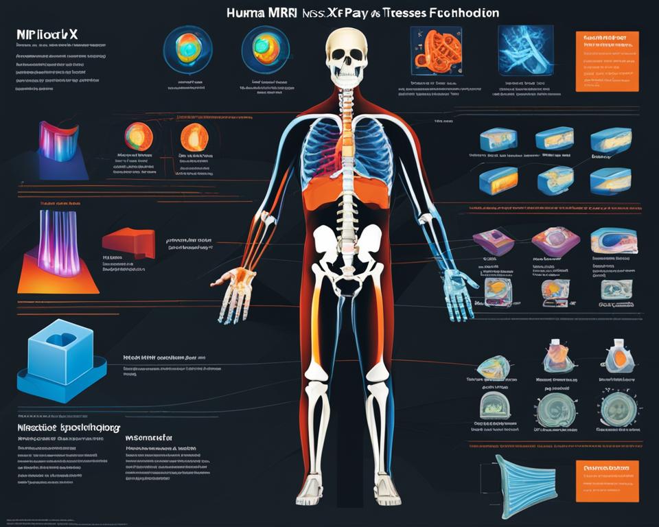
When it comes to diagnostic imaging, two common techniques used by doctors are MRI and X-ray. While both methods allow physicians to look inside the body and identify potential issues, there are key differences between the two.

Key Takeaways:
- MRI and X-ray are both diagnostic imaging techniques used to see inside the body.
- X-rays use radiation, while MRIs use a magnetic field and radio waves.
- X-rays are ideal for detecting fractures and dislocations, while MRIs are more effective at identifying soft tissue injuries and musculoskeletal conditions.
- X-rays are faster and more accessible, while MRIs provide greater detail and contrast resolution.
- Consult with your doctor to determine which imaging test is right for your specific condition.
What Is an X-Ray?
X-rays are the most common and widely available diagnostic imaging test used by doctors to visualize the internal structures of the body. They work by using radiation to create images. X-rays are quick and usually take only a few minutes to complete.
During an X-ray examination, the part of the body being imaged is positioned between the X-ray machine and the photographic film or digital X-ray sensor. The X-ray machine emits a small amount of radiation that passes through the body and is absorbed differently by different tissues. Dense objects such as bones appear white on the X-ray image, while less dense tissues such as muscles and organs appear shades of gray. X-rays are commonly used to diagnose and assess various conditions, including fractures, dislocations, tumors, infections, and bone degeneration.
“X-rays are a valuable tool in medicine, allowing doctors to see inside the body without the need for invasive procedures.”
Some common uses of X-rays include:
- Identifying broken bones and fractures
- Evaluating joint spaces and alignment
- Detecting tumors
- Determining the cause of lung problems like pneumonia
- Diagnosing dental issues like cavities and infections
X-rays are widely accessible and are often the first-line imaging tool due to their speed and lower cost compared to other imaging techniques. However, it’s important to note that X-rays use ionizing radiation, which carries potential risks. The benefits of X-rays generally outweigh the risks, especially when used judiciously by medical professionals.
| X-Ray Advantages | X-Ray Disadvantages |
|---|---|
|
|
What Is a CT Scan?
A CT scan, or computed tomography scan, is a more advanced form of X-ray that produces detailed, cross-sectional images of the body. It also uses radiation, but at a higher level of detail. CT scans are ideal for evaluating the size, shape, and position of bones, blood vessels, soft tissues, and organs. They can be used to diagnose a wide range of conditions such as appendicitis, cancer, trauma, heart disease, and musculoskeletal disorders.
During a CT scan, the patient lies on a table that slides into a machine resembling a large box. The machine rotates around the patient, taking many pictures from different angles to create a 3D image.
CT Scan vs X-ray:
While both CT scans and X-rays are imaging techniques, there are significant differences between the two. X-rays provide a more general overview of the body, while CT scans offer more detailed images that can reveal abnormalities in greater depth. CT scans are especially useful for identifying tumors, internal bleeding, and other complex conditions that may not be easily detected by X-rays. Additionally, CT scans can provide a three-dimensional view of the body, giving doctors a more comprehensive understanding of the patient’s condition.
Uses of CT Scan:
- Detecting and diagnosing diseases and injuries
- Evaluating the effectiveness of treatments and therapies
- Guiding biopsies and surgeries
- Planning radiation therapy and other medical procedures
- Screening for certain diseases, such as lung cancer
| CT Scan | X-ray | |
|---|---|---|
| Definition | A more advanced form of X-ray that produces detailed, cross-sectional images of the body | A diagnostic imaging technique that uses radiation to create images of the body’s internal structures |
| Uses | Identifying tumors, internal bleeding, complex conditions; guiding biopsies and surgeries; planning radiation therapy | Detecting fractures, dislocations, tumors, infections, and bone degeneration |
| Radiation | Uses a higher level of radiation | Uses a lower level of radiation |
Overall, CT scans are an essential tool in modern medicine, providing detailed images that can help doctors diagnose and treat various conditions. They offer a valuable alternative to X-rays when a more comprehensive view of the body is needed.
What Is an MRI?
An MRI, or magnetic resonance imaging, is a diagnostic imaging technique that uses a powerful magnet and radio waves to produce highly detailed images of the body’s structures. Unlike X-rays and CT scans, MRIs do not use radiation. MRIs are particularly effective at visualizing soft tissues, nerves, and blood vessels. They are often used to diagnose joint and bone problems, assess treatment progress, and evaluate brain abnormalities. MRIs offer excellent contrast resolution for both bones and soft tissues, making them especially useful for detecting cartilage loss, joint inflammation, nerve compression, and musculoskeletal injuries.
During an MRI, the patient lies on a table that slides into a cylindrical machine. The machine creates a magnetic field that causes the body’s tissues to resonate, and a computer translates this data into detailed images.
Compared to CT scans, MRIs provide higher resolution images, making them more suitable for diagnosing soft tissue injuries and abnormalities. MRIs are often recommended when X-rays or CT scans do not provide sufficient information or when a more detailed examination is needed. However, MRIs can take longer to complete, and some patients may experience discomfort or anxiety due to the confined space of the MRI machine.
MRI vs CT Scan
While CT scans and MRIs are both diagnostic imaging techniques, they have some important differences. CT scans use X-rays and provide detailed cross-sectional images of the body, making them particularly useful for evaluating bone structures and blood vessels. On the other hand, MRIs use magnets and radio waves to create highly detailed images of both bones and soft tissues.
CT scans are faster and more readily available compared to MRIs. They are often used in emergency situations or when a quick diagnosis is necessary. MRIs, on the other hand, offer better contrast resolution and are more effective at visualizing soft tissues, nerves, and blood vessels. They are often recommended for diagnosing conditions such as brain abnormalities, joint injuries, and musculoskeletal disorders.
| MRI | CT Scan | |
|---|---|---|
| Imaging Technique | Magnetic field and radio waves | X-rays |
| Radiation | No radiation | Uses radiation |
| Image Detail | High resolution, excellent contrast for soft tissues | Good resolution, excellent for bone and blood vessels |
| Availability | May not be available in all facilities | More readily available |
| Uses | Soft tissue injuries, abnormalities, joint and bone problems | Bone fractures, blood vessels, lung disorders |
Overall, the choice between an MRI and a CT scan depends on the specific condition being diagnosed and the information required by the healthcare provider. In some cases, both imaging techniques may be used together to provide a more comprehensive evaluation of the patient’s condition.
Similarities and Differences
When comparing MRI and X-ray, it is important to note both the similarities and differences between these two diagnostic imaging techniques. While both methods serve the purpose of providing insights into the body’s internal structures, they differ significantly in terms of their mechanisms and applications.
Similarities
Both MRI and X-ray are valuable tools used by medical professionals to aid in the diagnosis and treatment of various conditions. They provide important information about the body’s anatomy and help identify potential abnormalities or injuries. Additionally, both techniques are non-invasive, meaning they do not require any surgical procedures or interventions.
Differences
Despite the similarities, there are distinct differences between MRI and X-ray.
X-ray imaging uses radiation to create images of the body’s internal structures. It is particularly effective at detecting fractures, dislocations, and narrow joint spaces. In contrast, MRI utilizes a magnetic field and radio waves to produce highly detailed images of both bones and soft tissues. MRIs are more effective at identifying soft tissue injuries, inflammation, and musculoskeletal conditions.
| Comparison | X-ray | MRI |
|---|---|---|
| Imaging Mechanism | Radiation | Magnetic field and radio waves |
| Application | Fractures, dislocations, narrow joint spaces | Soft tissue injuries, inflammation, musculoskeletal conditions |
| Availability | Widely available | May not be available in all imaging facilities or emergency rooms |
Another notable difference is the availability of both imaging techniques. X-rays are widely accessible and often the first-line imaging tool, available in many healthcare facilities. On the other hand, MRIs may not be available in all imaging facilities or emergency rooms, limiting their accessibility.
In summary, while both MRI and X-ray play crucial roles in diagnosing and treating various medical conditions, they differ in their mechanisms, applications, and availability. It is essential to consult with your healthcare provider to determine which imaging test is most appropriate for your individual needs.
Conclusion
In conclusion, both MRI and X-ray are valuable diagnostic imaging techniques that play a crucial role in assessing and diagnosing various medical conditions. X-rays are faster and more accessible, making them ideal for quickly detecting fractures, dislocations, and narrow joint spaces. On the other hand, MRIs provide greater detail and contrast resolution, making them more effective at visualizing soft tissue injuries, inflammation, and musculoskeletal conditions.
When choosing between MRI and X-ray, it’s important to consider the specific condition and the information needed for an accurate diagnosis. X-rays are widely available and often the first-line imaging tool, especially for bone-related conditions. However, if the suspected condition involves soft tissues, nerves, or blood vessels, an MRI may be more appropriate. It’s always best to consult with your doctor to determine the most suitable imaging test for your individual situation.
In summary, while both MRI and X-ray have their advantages, the choice between them ultimately depends on the nature of the medical condition and the desired level of detail. Whether it’s identifying fractures or assessing soft tissue injuries, these imaging techniques offer valuable insights that enable doctors to provide appropriate treatment and care.
FAQ
What is the difference between an MRI and an X-ray?
An MRI uses a magnetic field and radio waves to create detailed images of the body’s structures, while an X-ray uses radiation to produce quick images of bones and internal structures.
What are X-rays used for?
X-rays are used to diagnose and assess conditions such as fractures, dislocations, tumors, infections, and bone degeneration.
What is a CT scan?
A CT scan, or computed tomography scan, is a more advanced form of X-ray that produces detailed, cross-sectional images of the body. It is ideal for evaluating bones, blood vessels, soft tissues, and organs.
What is an MRI used for?
An MRI is used to visualize soft tissues, nerves, and blood vessels. It is often used to diagnose joint and bone problems, assess treatment progress, and evaluate brain abnormalities.
Are X-rays and MRIs widely available?
X-rays are the most common and widely available diagnostic imaging test. MRIs may not be available in all imaging facilities or emergency rooms.
Which imaging technique is better for detecting fractures?
X-rays are ideal for detecting fractures and dislocations, as they provide quick images of bones.
Which imaging technique is better for identifying soft tissue injuries?
MRIs are more effective at identifying soft tissue injuries, inflammation, and musculoskeletal conditions.
How long does an X-ray usually take?
X-rays are quick and usually take only a few minutes to complete.
How does an MRI work?
During an MRI, the patient lies on a table that slides into a cylindrical machine. A powerful magnet and radio waves create detailed images of the body’s structures.
How does a CT scan work?
During a CT scan, the patient lies on a table that slides into a machine resembling a large box. The machine rotates around the patient, taking many pictures from different angles to create a 3D image.
Which imaging test should I choose for my specific condition?
It is important to consult with your doctor to determine the most appropriate imaging test for your individual situation.

