When it comes to medical procedures, understanding the difference between a PET scan and a CT scan can make a world of difference. These two diagnostic tools serve unique purposes and utilize different technologies. Let’s dive into the details and explore the contrasts between these two essential medical procedures.

Key Takeaways:
- PET scans and CT scans are both important medical procedures used for diagnosis.
- A CT scan creates detailed non-moving images of organs, bones, and tissues, providing valuable anatomical information.
- PET scans show how the tissues in the body function on a cellular level, revealing metabolic activity.
- CT scans use x-rays, while PET scans use radioactive materials that emit energy.
- Both procedures are accurate, painless, and noninvasive, providing valuable diagnostic information.
What is a CT Scan?
A CT scan, or computed tomography, is an imaging procedure that uses x-rays and computer technology to create a detailed 3-D image of the inside of the body. It is a non-invasive procedure commonly used to check for abnormalities in various parts of the body, such as the brain, spine, neck, chest, or abdomen. CT scans provide valuable information about both hard tissues, like bones, and soft tissues, like muscles and organs.
A CT scan works by using multiple x-ray beams to capture images of the body from different angles. These images are then translated into cross-sectional pictures on a computer monitor. The detailed 3-D image created by a CT scan allows doctors to visualize the internal structures of the body and identify any abnormalities or conditions that may be present.
CT scans are highly effective in diagnosing a wide range of conditions and are often used in emergency situations due to their speed and accuracy. They are quick and painless for patients, making them a valuable tool in the field of medical imaging.
How Does a CT Scan Work?
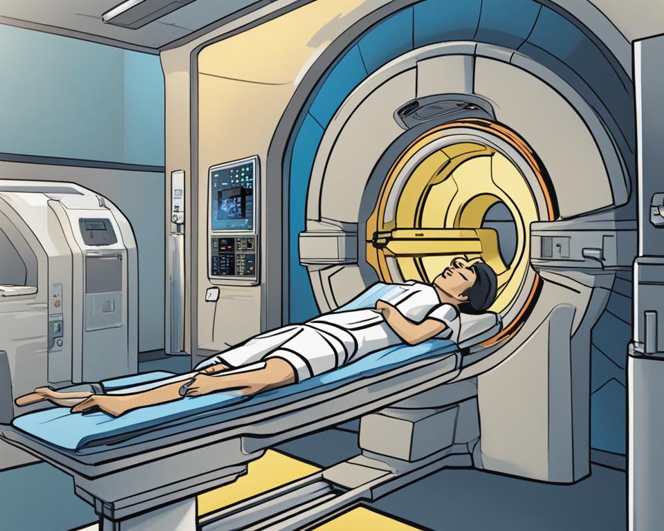
During a CT scan, x-ray beams are used to capture images of the body from different angles. The patient lies on an exam table and is moved through a CT scan machine. The machine emits multiple x-ray beams, which pass through the body and are detected by sensors on the other side. These detected signals are then reconstructed by a computer to create cross-sectional pictures of the body. These images provide detailed information about the internal structures, such as organs, bones, and tissues.
To enhance the visibility of certain areas, contrast material may be administered to the patient. This material can be in the form of an oral drink, injection, or enema, depending on the area of the body being scanned. The contrast material helps highlight specific structures or abnormalities, making them easier to detect and analyze. The entire CT scan procedure is quick and painless, typically taking only a few minutes to complete.
“CT scans are a valuable diagnostic tool due to their ability to provide detailed, cross-sectional images of the body. This enables healthcare professionals to accurately identify and evaluate various conditions, such as tumors, fractures, infections, and more. The non-invasive nature of CT scans makes them a preferred choice for doctors and patients alike, as they carry a relatively low risk and do not require any surgical procedures.”
Advantages of CT Scans:
- Provide detailed images of internal structures
- Quick and painless procedure
- Can be performed with or without contrast material
- Accurate detection and evaluation of various conditions
- Non-invasive and low-risk
Disadvantages of CT Scans:
- Exposure to ionizing radiation (although minimal)
- Not recommended for pregnant women due to radiation exposure
- Possible allergic reactions to contrast material
| CT Scan | PET Scan |
|---|---|
| Produces detailed, cross-sectional images | Shows metabolic activity in tissues and organs |
| Uses x-ray beams | Uses radioactive tracers |
| Painless and quick procedure | Takes longer and requires waiting period for tracer uptake |
| No residual radiation in the body | Small amount of residual radiation may remain |
What is a PET Scan?
A PET scan, or positron emission tomography, is a form of nuclear medicine imaging. It involves the injection of a tiny amount of radioactive material, known as a radiotracer, into the patient’s vein. The radiotracer moves through the body and collects in tissues and organs, allowing the scan to show how these tissues and organs are functioning. PET scans are commonly used to detect or monitor cancer and assess heart and brain functioning.
During a PET scan, the radiotracer emits positrons, which are positively charged particles. When the positrons collide with electrons in the body, they annihilate each other, producing gamma rays. The PET scanner detects these gamma rays and uses them to create 3-D images of the body’s internal structures and metabolic activity.
PET scans are particularly valuable in assessing the function of tissues and organs because they provide information on a cellular level. This makes them useful for diagnosing and staging cancer, evaluating heart function, and assessing brain activity and neurological disorders. PET scans can also be used to monitor the effectiveness of cancer treatments and guide targeted therapies.
| Advantages of PET Scans | Limitations of PET Scans |
|---|---|
|
|
Overall, PET scans offer valuable insights into tissue and organ functioning, making them a crucial tool in diagnosing and monitoring various medical conditions. While there are limitations to their use, the benefits outweigh the drawbacks in many cases, especially when detailed functional information is required.
How Does a PET Scan Work?
A PET scan, or positron emission tomography, is a medical imaging procedure that utilizes a radioactive tracer to create detailed 3-D images of the body’s tissues and organs. This non-invasive test helps doctors assess the functioning of various body systems and detect abnormalities.
After the injection of a radioactive tracer, which is usually a small amount of a glucose compound, the patient waits for the tracer to be absorbed by the body. The radioactive tracer emits energy in the form of positrons, which are detected by the PET scanner. The scanner captures the energy signals and converts them into visual 3-D images on a computer monitor.
The PET images reveal metabolic activity in the body, indicating how well tissues and organs are functioning. Areas with high metabolic activity, such as cancer cells, appear as bright spots on the images, while areas with low activity appear darker. By analyzing these images, doctors can diagnose and monitor various conditions, including cancer, brain disorders, and heart disease.
Advantages of PET Scans:
- Ability to detect diseases at an early stage by revealing molecular activity
- Non-invasive and painless procedure
- Provides detailed 3-D images of tissues and organs
- Helps evaluate treatment effectiveness and disease progression
When combined with a CT scan, PET scans can provide more comprehensive and precise information. This hybrid imaging technique, known as PET/CT, allows for the visualization of both metabolic activity and the anatomical structure of the body. PET/CT scans are particularly valuable in oncology, as they can help accurately stage cancer and guide treatment decisions.
Table: Comparison of PET Scans and CT Scans
| Aspect | PET Scan | CT Scan |
|---|---|---|
| Focus | Reveals metabolic activity | Provides anatomical information |
| Technology | Uses radioactive tracers | Uses x-rays |
| Procedure Time | Takes longer due to tracer absorption | Relatively quick |
| Radiation | Leaves residual radiation in the body | No residual radiation |
The Difference Between a CT Scan and a PET Scan
When it comes to medical imaging, CT scans and PET scans are two commonly used procedures with distinct differences. CT scans, short for computed tomography, create detailed non-moving images of bones, organs, and tissues, providing valuable anatomical information. On the other hand, PET scans, or positron emission tomography, reveal how the tissues in the body function on a cellular level, showing metabolic activity. CT scans use x-rays, while PET scans use radioactive materials that emit energy.
Another difference between the two procedures is the time it takes to complete them. CT scans are relatively quick, whereas PET scans can be more time-consuming, depending on the patient’s condition. Additionally, after a CT scan, there is no residual radiation in the body, while a small amount of radiation may remain after a PET scan.
One area where PET scans shine is in the detection of cancer. Due to their ability to show molecular activity, PET scans are highly reliable for detecting cancer at early stages. This makes them an invaluable tool in cancer diagnosis and monitoring.
In summary, while CT scans provide detailed anatomical information, PET scans reveal metabolic activity. Understanding these differences can help patients and doctors determine which imaging procedure is most appropriate for their specific needs.
| CT Scan | PET Scan |
|---|---|
| Creates detailed non-moving images of organs, bones, and tissues | Reveals how tissues function on a cellular level |
| Uses x-rays for imaging | Uses radioactive materials that emit energy |
| Relatively quick procedure | Can be more time-consuming |
| No residual radiation after the procedure | Small amount of radiation may remain |
| Provides valuable anatomical information | Highly reliable for detecting cancer |
Similarities Between CT Scans and PET Scans
While CT scans and PET scans have distinct differences, they also share several important similarities. These imaging procedures are both commonly performed on an outpatient basis, meaning they do not require a hospital stay. This makes them convenient and accessible for patients seeking diagnostic information about their health.
Both CT scans and PET scans are highly accurate in detecting cancer and other medical conditions. These scans provide detailed and valuable information that helps doctors make accurate diagnoses and develop appropriate treatment plans for their patients. By using advanced technology and capturing images of the body’s internal structures, CT scans and PET scans play a crucial role in identifying abnormalities and monitoring disease progression.
Additionally, both CT scans and PET scans are noninvasive and painless procedures. This means that patients can undergo these imaging tests without the need for surgery or any discomfort. These characteristics make CT scans and PET scans accessible to a wide range of patients, including those with medical conditions that may make invasive procedures risky or undesirable.
Overall, CT scans and PET scans are essential tools in modern medicine, allowing doctors to diagnose symptoms accurately and provide appropriate medical care. By understanding the similarities and differences between these two imaging procedures, patients and healthcare professionals can make informed decisions about the most appropriate diagnostic approach for individual cases.
| Similarities Between CT Scans and PET Scans |
|---|
| Both can be performed on an outpatient basis |
| Both are accurate in detecting cancer and other conditions |
| Both are noninvasive and painless procedures |
Conclusion
Understanding the PET scan and CT scan is essential for making informed healthcare decisions. These diagnostic procedures play crucial roles in identifying and diagnosing various medical conditions. By recognizing the differences between PET scans and CT scans, patients and doctors can work together to determine the most appropriate course of action.
A PET scan provides valuable insight into the cellular functioning of tissues and organs, allowing for the detection and monitoring of conditions such as cancer. On the other hand, a CT scan offers detailed images of bones, organs, and tissues, aiding in the identification of anatomical abnormalities. While both scans are integral to modern medicine, they utilize different technologies and approaches.
By understanding these differences, patients can actively participate in their healthcare journey. They can discuss with their doctors the advantages and limitations of each scan, and together, make informed decisions about which approach best suits their needs. Whether it’s ensuring early cancer detection or obtaining detailed anatomical information, the use of PET scans and CT scans can lead to more accurate diagnoses, effective treatments, and improved health outcomes.
Ultimately, the understanding of PET scans and CT scans empowers individuals to be proactive in managing their health. It enables them to have informed discussions with their healthcare providers, weigh the benefits and risks, and actively participate in the decision-making process. By taking an active role in their healthcare decisions, individuals can work towards better health and well-being.
FAQ
What is a CT Scan?
A CT scan, or computed tomography, is a non-invasive imaging procedure that uses x-rays and computer technology to create a detailed 3-D image of the inside of the body.
How does a CT Scan work?
During a CT scan, the patient lies on an exam table and is moved through a CT scan machine. The machine uses multiple x-ray beams to capture images of the body from different angles, which are then translated into cross-sectional pictures on a computer monitor.
What is a PET Scan?
A PET scan, or positron emission tomography, is a form of nuclear medicine imaging. It involves the injection of a tiny amount of radioactive material, known as a radiotracer, into the patient’s vein. The radiotracer moves through the body and collects in tissues and organs, allowing the scan to show how these tissues and organs are functioning.
How does a PET Scan work?
After the injection of the radioactive tracer, the patient waits for the body to absorb the tracer, usually for about an hour. Then, the patient lies on an exam table that slides into a PET scanner. The scanner detects the tracer and converts the data into 3-D images on a computer monitor.
What is the difference between a CT Scan and a PET Scan?
The main difference between a CT scan and a PET scan lies in their focus and the materials they use. A CT scan creates detailed non-moving images of organs, bones, and tissues, providing valuable anatomical information. On the other hand, a PET scan shows how the tissues in the body function on a cellular level, revealing metabolic activity. CT scans use x-rays, while PET scans use radioactive materials that emit energy. A CT scan is a relatively quick procedure, while a PET scan can take longer depending on the patient’s condition. After a CT scan, there is no residual radiation in the body, but a small amount of radiation may remain after a PET scan. Additionally, PET scans are highly reliable for detecting cancer at early stages due to their ability to show molecular activity.
What are the similarities between CT scans and PET scans?
Despite their differences, CT scans and PET scans share several similarities. Both procedures can be performed on an outpatient basis, meaning they do not require a hospital stay. They are both accurate, painless, and noninvasive, providing valuable diagnostic information. They can both be used to detect cancer and help eliminate the need for exploratory surgery. Most importantly, both CT scans and PET scans enable doctors to diagnose the cause of a patient’s symptoms, leading to effective treatment and better health outcomes.
Conclusion
Understanding the difference between a PET scan and a CT scan is crucial for making informed healthcare decisions. While both procedures serve important diagnostic purposes, they focus on different aspects of the body and use different technologies. By understanding these differences and similarities, patients and doctors can work together to ensure the best possible care and outcomes.

![Ray Dalio Quotes [Principles, Life, Investment]](https://tagvault.org/wp-content/uploads/2023/04/Screen-Shot-2023-04-19-at-7.57.49-PM.png)