Welcome to our comprehensive guide on the key differences between myofibrils and muscle fibers. As a beginner in the world of muscles, you may have come across these terms and wondered what sets them apart. In this article, we will delve into their functions, structures, compositions, and more. So, let’s get started!
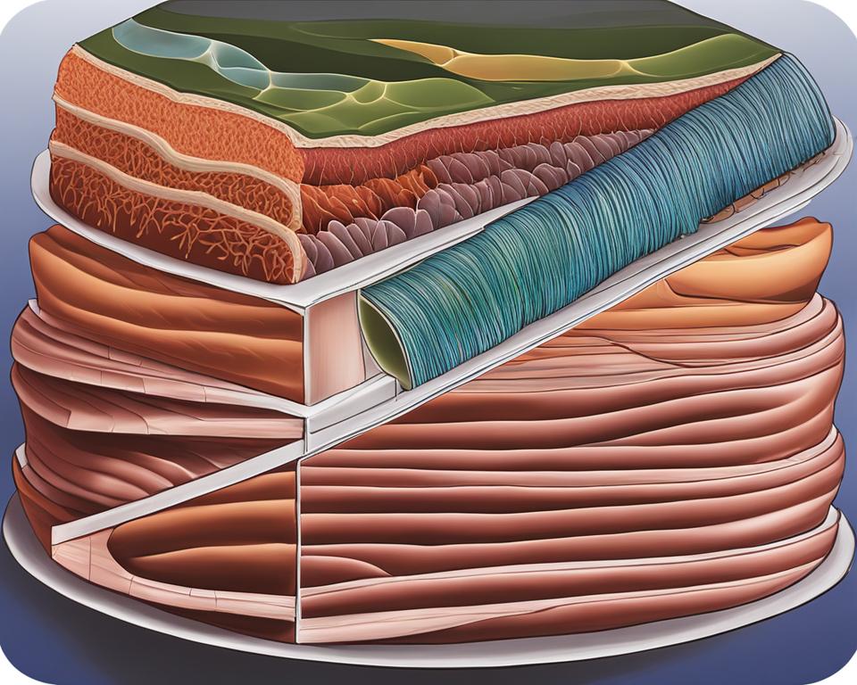
But first, let’s clarify the basics. Muscle fibers, also known as myocytes, are the tubular cells that make up muscle tissues. They are long bundles of cells that play a crucial role in muscle contraction. On the other hand, myofibrils are the rod-like units that form the basic structure of muscle fibers. They are composed of various proteins that are responsible for muscle contractions.
Now that we have a basic understanding of the terms, let’s explore them further.

- Myofibrils and muscle fibers are essential components of the muscular system.
- Myofibrils are the basic units of muscle fibers, while muscle fibers are tubular cells.
- Myofibrils are composed of various proteins responsible for muscle contractions.
- Muscle fibers are long bundles of cells that enable muscle contraction.
- Understanding the differences between myofibrils and muscle fibers enhances our knowledge of muscle function and anatomy.
Structure of Muscle Fibers
Skeletal muscles are composed of long, cylindrical bundles of cells called muscle fibers. These muscle fibers are encased in a protective connective tissue called endomysium and organized into groups called fascicles. Within each muscle fiber, there are thousands of myofibrils, which are the functional units responsible for muscle contraction.
The structure of muscle fibers is designed to support their function. Each muscle fiber contains numerous myofibrils, which are composed of thick and thin filaments made up of proteins called myosin and actin, respectively. These myofilaments are organized into repeating units called sarcomeres, which give muscle fibers their striated appearance. The arrangement of these sarcomeres allows for coordinated muscle contraction.
The anatomy of a muscle fiber also includes various organelles and structures that contribute to its function. The sarcoplasmic reticulum is a specialized type of endoplasmic reticulum that stores and releases calcium ions, which are crucial for muscle contraction. T-tubules are invaginations of the muscle fiber membrane that allow for the rapid transmission of electrical signals necessary for muscle contraction. Additionally, mitochondria, the energy powerhouses of cells, are abundant in muscle fibers to provide the ATP needed for muscle function.
Table: Components of Muscle Fiber Structure
| Component | Description |
|---|---|
| Myofibrils | Long, rod-like structures composed of myosin and actin proteins responsible for muscle contraction. |
| Sarcomeres | Repeating units of myofibrils that give muscle fibers their striated appearance and enable coordinated muscle contraction. |
| Sarcoplasmic Reticulum | Specialized endoplasmic reticulum that stores and releases calcium ions, crucial for muscle contraction. |
| T-tubules | Invaginations of the muscle fiber membrane that transmit electrical signals for muscle contraction. |
| Mitochondria | Organelles that produce ATP, the energy molecule required for muscle function. |
In summary, muscle fibers have a complex structure that enables their function. They are composed of thousands of myofibrils, which contain thick and thin filaments arranged in sarcomeres. Various organelles, such as the sarcoplasmic reticulum and mitochondria, contribute to the contraction and energy production of muscle fibers.
Types of Muscle Fibers
When it comes to muscle fibers, there are three main types: slow twitch fibers, fast oxidative fibers, and fast glycolytic fibers. Each type has distinct characteristics and plays a specific role in muscle function.
Slow twitch fibers, also known as Type I fibers or red slow fibers, are designed for endurance activities. They have a high capacity for aerobic metabolism, meaning they can generate energy efficiently using oxygen. Slow twitch fibers are rich in mitochondria, which fuel their endurance capabilities. These fibers are useful for activities such as long-distance running or cycling.
In contrast, fast twitch fibers can be further divided into two subtypes: fast oxidative fibers (Type IIa fibers or red fast fibers) and fast glycolytic fibers (Type IIb fibers or white fast fibers). Fast oxidative fibers have intermediate characteristics between slow and fast glycolytic fibers. They have a high capacity for aerobic metabolism and can also generate energy anaerobically when needed. Fast glycolytic fibers, on the other hand, rely primarily on anaerobic metabolism for energy production. These fibers are well-suited for short bursts of intense activity, such as weightlifting or sprinting.
| Type of Muscle Fiber | Color | Metabolic Capacity | Function |
|---|---|---|---|
| Slow Twitch (Type I) Fibers | Red | High aerobic capacity | Endurance activities |
| Fast Oxidative (Type IIa) Fibers | Red | Intermediate | Moderate-intensity exercises |
| Fast Glycolytic (Type IIb) Fibers | White | High anaerobic capacity | Explosive, high-intensity exercises |
Each type of muscle fiber has its own unique characteristics and functions, allowing our muscles to perform a wide range of activities. Whether it’s the endurance of slow twitch fibers, the versatility of fast oxidative fibers, or the power of fast glycolytic fibers, our muscles work together to help us excel in various physical endeavors.
The Role of Muscle Fiber Types in Physical Performance
The distribution of muscle fiber types in an individual’s body can influence their athletic performance. For example, individuals with a higher proportion of slow twitch fibers may excel in endurance activities, while those with a higher proportion of fast twitch fibers may excel in explosive and high-intensity activities.
It’s important to note that muscle fiber types are not fixed and can be influenced by various factors, including training. Endurance training, such as long-distance running, can lead to an increase in the proportion of slow twitch fibers, enhancing an individual’s endurance capabilities. On the other hand, resistance training, such as weightlifting, can lead to an increase in the proportion of fast twitch fibers, improving an individual’s strength and power.
In summary, understanding the different types of muscle fibers and their unique characteristics can help individuals tailor their training and maximize their performance in specific activities. By focusing on the appropriate training methods, individuals can optimize the development and function of their muscles, ultimately enhancing their overall physical performance.
Myofibril: The Basic Unit of Muscle Fiber
The myofibril is a vital component of muscle fibers, serving as the fundamental unit for muscle contraction and function. Composed primarily of actin and myosin proteins, myofibrils play a crucial role in generating the force required for muscle movement.
The structure of a myofibril consists of repeating sections known as sarcomeres, which give skeletal muscle fibers their striated appearance. Within each sarcomere, thick filaments of myosin and thin filaments of actin are organized in a highly ordered fashion. This arrangement allows for the sliding motion of these filaments during muscle contraction.
Myofibrils are organized into distinct bands and zones, each contributing to their overall composition and organization. The Z-lines delineate the boundaries of individual sarcomeres, while the A-band represents the region of overlapping thick and thin filaments. The I-band corresponds to the region where only thin filaments are present, and the H-zone represents the area within the A-band where only thick filaments are present.
Overall, the myofibril’s composition, organization, and structure facilitate the coordinated contraction of muscle fibers, enabling various types of muscle movements in the body.
Table: Myofibril Composition and Organization
| Component | Description |
|---|---|
| Actin | Main protein component of thin filaments |
| Myosin | Main protein component of thick filaments |
| Z-lines | Boundary lines of individual sarcomeres |
| A-band | Region of overlapping thick and thin filaments |
| I-band | Region where only thin filaments are present |
| H-zone | Region within the A-band where only thick filaments are present |
Similarities Between Myofibril and Muscle Fiber
When comparing myofibrils and muscle fibers, several similarities can be identified in terms of their shape, arrangement, and function. These similarities contribute to the overall functioning of the muscular system.
Shape and Arrangement
Both myofibrils and muscle fibers share a tubular shape. Myofibrils are elongated cylindrical structures, while muscle fibers are the individual tubular cells that make up the muscle tissue. This tubular shape allows for efficient transmission of signals and forces along the length of the muscle fiber, enabling coordinated movement.
In terms of arrangement, both myofibrils and muscle fibers are organized in parallel within the muscle. This parallel arrangement enables synchronized contraction and relaxation of muscle fibers, leading to efficient muscle movement. The parallel arrangement also allows for the distribution of forces throughout the muscle, resulting in smooth and coordinated movements.
Function
Both myofibrils and muscle fibers play crucial roles in muscle contraction. Myofibrils consist of thin and thick filaments that slide past each other during contraction, resulting in the shortening of the muscle fiber. This sliding filament mechanism is common to all muscle fibers and myofibrils, regardless of their type.
Additionally, both myofibrils and muscle fibers are involved in force production. As myofibrils contract, they generate tension that is transmitted to the surrounding connective tissues and eventually to the muscle fibers. This tension allows for the movement of bones and joints, facilitating various body movements.
In summary, myofibrils and muscle fibers share similarities in terms of their tubular shape, parallel arrangement, and involvement in muscle contraction and force production. These similarities contribute to the overall functioning of the muscular system, enabling coordinated movement and efficient force generation.
The Relationship Between Myofibril and Muscle Fiber
In order to understand the relationship between myofibrils and muscle fibers, it’s important to first grasp their individual functions and structures. Myofibrils, as mentioned earlier, are the basic rod-like units within a muscle fiber. These cylindrical organelles are composed of actin and myosin proteins, along with various other types of proteins. They are organized into thick and thin filaments called myofilaments, which play a crucial role in muscle contraction.
On the other hand, muscle fibers are the tubular cells that make up the muscular tissue. Each muscle fiber contains numerous myofibrils, making them the building blocks of muscle fibers. The myofibrils within a muscle fiber are responsible for generating the force required for muscle contraction. This complex relationship between myofibrils and muscle fibers allows for the coordinated and efficient functioning of skeletal muscles.
It’s important to note that while myofibrils are the structural units of muscle fibers, muscle fibers themselves are responsible for producing the actual contractions that enable movement. The myofibrils within a muscle fiber work together to generate force and shorten the length of the muscle fiber, resulting in muscle contraction. This connection between myofibrils and muscle fibers highlights the intricate and interdependent nature of the muscular system.
Table: Comparing Myofibrils and Muscle Fibers
| Aspect | Myofibrils | Muscle Fibers |
|---|---|---|
| Definition | The basic rod-like units within a muscle fiber | The tubular cells that make up the muscular tissue |
| Composition | Composed of actin, myosin, and other proteins | Composed of bundles of myofibrils |
| Function | Generate force and enable muscle contraction | Produce contractions for muscle movement |
| Interdependence | Essential structural units of muscle fibers | Contain hundreds of myofibrils |
This table provides a clear comparison between myofibrils and muscle fibers, showcasing their distinct characteristics and interdependence. While myofibrils are the building blocks and functional units within muscle fibers, muscle fibers themselves are responsible for generating the actual contractions needed for movement.
By understanding the relationship between myofibrils and muscle fibers, we can gain a deeper appreciation for the complexity and efficiency of the muscular system. The coordinated interaction between these two components allows for the remarkable capabilities of skeletal muscles, enabling us to perform a wide range of movements and activities.
Difference Between Myofibril and Muscle Fiber
When it comes to understanding the muscular system, it is important to distinguish between myofibrils and muscle fibers. While these terms may sound similar, they refer to distinct components of the muscle. Let’s explore the key differences between myofibrils and muscle fibers.
Composition and Structure:
Myofibrils are rod-like structures that make up the basic unit of a muscle fiber. They consist of thin and thick filaments, primarily composed of actin and myosin proteins, respectively. The arrangement of these filaments within the myofibrils allows for muscle contraction. On the other hand, muscle fibers refer to the tubular cells that form the whole muscle. Each muscle fiber contains numerous myofibrils, which are responsible for the contraction and relaxation of the muscle.
Function:
Myofibrils play a crucial role in muscle contraction. The interaction between actin and myosin filaments shortens the length of the myofibrils, resulting in the muscle fiber contracting. This contraction generates the force required for movement. Muscle fibers, on the other hand, are responsible for transmitting the signals from the nervous system to the myofibrils, enabling coordinated muscle contractions.
Summary:
In summary, myofibrils are the structural units within a muscle fiber, composed of actin and myosin filaments. In contrast, muscle fibers are the tubular cells that contain multiple myofibrils. While myofibrils facilitate muscle contraction, muscle fibers transmit signals and coordinate muscle movements. Understanding the difference between these two components is essential for comprehending how muscles function.
| Aspect | Myofibril | Muscle Fiber |
|---|---|---|
| Composition | Primarily actin and myosin proteins | Tubular cells forming the muscle |
| Structure | Rod-like units within a muscle fiber | Tubular cells that make up the whole muscle |
| Function | Responsible for muscle contraction | Transmits signals and coordinates muscle movements |
Conclusion
In conclusion, understanding the differences between myofibrils and muscle fibers is crucial for comprehending the intricate workings of the muscular system. Myofibrils serve as the fundamental building blocks within muscle fibers, while muscle fibers are the tubular cells responsible for muscle contractions.
Myofibrils are composed of thin and thick filaments, and their organization into sarcomeres gives muscle fibers their distinct striated appearance. On the other hand, muscle fibers consist of numerous myofibrils that work together to generate the force necessary for movement.
By recognizing the similarities and disparities between myofibrils and muscle fibers, we can gain a deeper comprehension of muscle function and performance. Their interdependent relationship demonstrates how these vital components collaborate to facilitate muscle contraction, enabling us to perform various physical activities.
FAQ
What is the difference between myofibril and muscle fiber?
The main difference is that myofibril is the basic unit of a muscle fiber, while a muscle fiber refers to the tubular cells of the muscle. Myofibrils are composed of thin and thick filaments, while muscle fibers are composed of numerous myofibrils.
What are the types of muscle fibers?
There are three main types of muscle fibers: type I fibers (slow twitch fibers or red slow fibers), type IIa fibers (fast oxidative fibers or red fast fibers), and type IIb fibers (fast glycolytic fibers or white fast fibers). Each type has different characteristics in terms of function and performance.
What is the function of myofibrils?
Myofibrils are responsible for muscle contraction. They are composed mainly of actin and myosin proteins, along with other types of proteins. Myofibrils are organized into thick and thin filaments called myofilaments, which are responsible for muscle contraction.
What is the structure of muscle fibers?
Muscle fibers are composed of long bundles of cells called muscle fibers or myocytes. They are protected by a connective tissue called epimysium and are organized into fascicles. Each muscle fiber contains a large number of myofibrils, which are bundles of myosin and actin proteins responsible for muscle contraction.
How are myofibrils and muscle fibers similar?
Both myofibrils and muscle fibers are responsible for muscle contractions. They are tubular in shape and arranged in parallel inside the muscle. Myofibrils are the basic units of muscle fibers, and each muscle fiber contains hundreds of myofibrils, which are essential for muscle contraction.
What is the relationship between myofibril and muscle fiber?
Myofibrils make up the structural units of muscle fibers. They are cylindrical organelles composed mainly of actin and myosin proteins. Muscle fibers, on the other hand, are the tubular cells of the muscle that contain multiple myofibrils. Together, myofibrils and muscle fibers work together to enable muscle contraction.
What are the differences between myofibril and muscle fiber?
The main difference is that myofibril is the basic rod-like unit of a muscle fiber, while a muscle fiber refers to the tubular cells of the muscle. Myofibrils are composed of thin and thick filaments, while muscle fibers are composed of numerous myofibrils. Additionally, myofibrils are organized into sarcomeres, which give muscle fibers their striated appearance.

![Ray Dalio Quotes [Principles, Life, Investment]](https://tagvault.org/wp-content/uploads/2023/04/Screen-Shot-2023-04-19-at-7.57.49-PM.png)